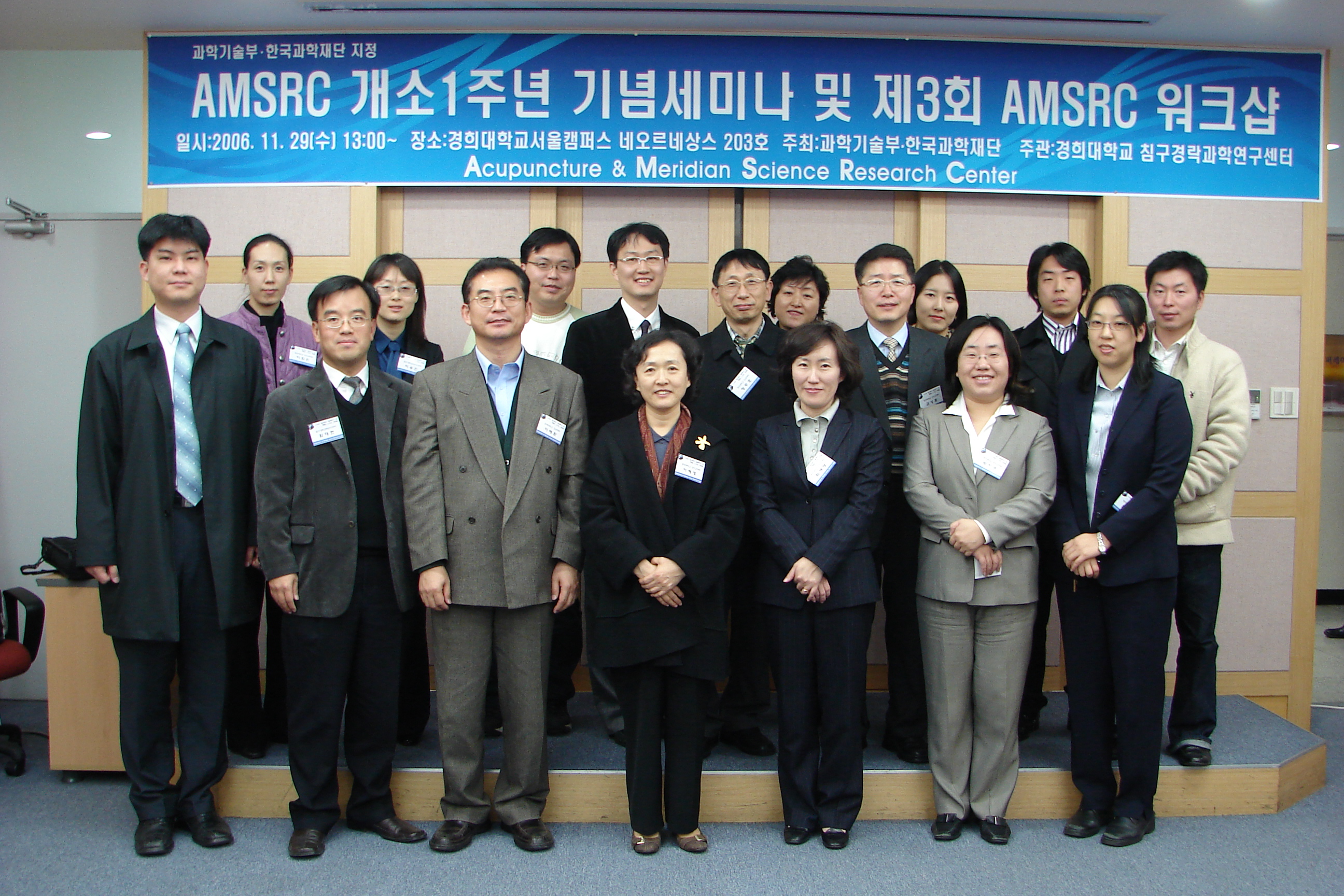
침구경락과학연구센터 개소1주년 기념세미나
> 연사 : 오 태 환 교수
> 소속 : 노인성및뇌질환연구센터
> 주제 : Therapeutic Interventions for Acute Spinal Cord Injury
> 일시 : 2005. 11. 29(수요일) p.m 13:00 ~
> 장소 : 경희대(서울) 네오르네상스 203호
> 주최 : 과학기술부. 한국과학재단
> 주관 : 경희대학교 침구경락과학연구센터
> Abstract :
Therapeutic Interventions for Acute Spinal Cord Injury
Tae Hwan Oh
Age-Related and Brain Diseases Research Center,
Kyung Hee University, Seoul 130-701, Korea
Traumatic spinal cord injury (SCI) initiates a complex series of cellular and molecular events that induce massive apoptotic cell death leading to permanent neurological deficits in human. However, the external apoptotic death signal(s) generated after SCI has not been identified. Furthermore, there is still no therapeutic agent effective for recovering motor functions after SCI in humans. Only one agent, methylprednisolone (MP), is currently being applied clinically to treat SCI. However, the clinical significance of recovery after MP treatment is unclear and must be considered in the light of potential adverse effects of its high-dose treatment. Although a number of potential pharmacological treatments have been investigated, other therapeutic agents designed to reduce cell death after SCI must be investigated.
Previously we reported that TNF-αis served as an external signal initiating apoptosis in neurons and oligodendrocytes after SCI and the apoptotic cascade initiated by TNF-α may be mediated in part by NO via up-regulation of iNOS induced in response to TNF-α. We also found that anti-inflammatory drug, minocycline and 17-estradiol reduce apoptosis and improve functional recovery after SCI. Rats received a mild, weight-drop contusion injury to the spinal cord and were treated with the vehicle or drugs. Using the Basso-Beattie-Bresnahan (BBB) locomotor open field behavioral rating test, we found that BBB scores were significantly improved in drug-treated rats as compared with those in vehicle-treated rats after SCI.In response to treatment with drugs, the lesion size was also significantly reduced after SCI. Furthermore, drug treatment significantly reduced the number of TUNEL-positive cells and DNA laddering after SCI as compared to that of the vehicle control. RT-PCR analyses revealed that minocycline treatment increased expression of interleukin-10 mRNA but decreased TNF-α expression. Recently we also found that minocycline improves functional recovery after SCI in part by reducing apoptosis of oligodendrocytes via inhibition of proNGF production in microglia. Especially, p38MAPK was only activated in microglia, and minocycline treatment inhibited proNGF production by inhibition p38MAPK activation after SCI. Furthermore, minocycline treatment significantly inhibited p75NTR expression and p75NTR-mediated apoptosis of oligodendrocytes, leading to inhibition of demyelination and axon degeneration as compared with vehicle control. We also found that 17β-estradiol significantly increases the expression of the anti-apoptotic genes, bcl-2 and bcl-x at early time and up-regulates ERK and AKT phosphorylation, which play as cell survival factors after SCI. In addition, 17β-estradiol treatment attenuated JNK activation at delayed time after injury, which is involved in apoptosis of oligodendrocytes, leading to functional improvement. These data suggest that after SCI, minocycline or 17β-estradiol treatment improved functional recovery in injured rat in part by regulating expression of cytokines, proNGF or anti-apoptotic genes, respectively, ultimately reducing apoptotic cell death. The significance of the proposed research is that it could lead us to therapeutic interventions for preventing cell death thereby improving functional recovery after SCI. This research was supported in part by the Korea MOST Neurobiology Research Program and NIH (US).
